※Alphabetical order
| Speaker | Dr. Pablo A. Iglesias (The Johns Hopkins University) |
| Title | Theoretical and Experimental Analysis of Chemotactic Systems in Biology |
| Abstract | Many biological systems have the ability to sense the direction of external chemical sources and respond by polarizing and migrating toward chemoattractants or away from chemorepellants. This phenomenon, referred to as chemotaxis, is crucial for proper functioning of single cell organisms, such as bacteria and amoebae, as well as multi-cellular systems as complex as the immune and nervous systems. Chemotaxis also appears to be important in wound healing and tumor metastasis. I will discuss our groups efforts at elucidating the mechanisms underlying chemotaxis. Using known biochemical data, we have developed mathematical models that can account for many of the observed chemotactic behavior of the model organism Dictyostelium. I will discuss experiments used to test these models. Finally, I will describe how information-theoretic methods can be used to evaluate the optimality of the gradient sensing mechanisms. |
| References | * Levchenko A, Iglesias PA: *Models of eukaryotic gradient sensing: application to chemotaxis of amoebae and neutrophils*. /Biophys J /2002, *82*:50-63. * Janetopoulos C, Ma L, Devreotes PN, Iglesias PA: *Chemoattractant-induced phosphatidylinositol 3,4,5-trisphosphate accumulation is spatially amplified and adapts, independent of the actin cytoskeleton*. /Proc Natl Acad Sci U S A /2004, *101*:8951-8956. * Ma L, Janetopoulos C, Yang L, Devreotes PN, Iglesias PA: *Two complementary, local excitation, global inhibition mechanisms acting in parallel can explain the chemoattractant-induced regulation of PI(3,4,5)P3 response in dictyostelium cells*. /Biophys J /2004, *87*:3764-3774. * Franca-Koh J, Kamimura Y, Devreotes P: *Navigating signaling networks: chemotaxis in Dictyostelium discoideum*. /Curr Opin Genet Dev /2006, *16*:333-338. |
| Speaker | 見学美根子 (理化学研究所) |
| Title | Mechanisms of neuronal migration |
| Abstract | Newborn neurons in
the developing brain migrate from the site of birth to their
destination for integration into specific neural circuits.
Neuronal migration is highly directional movement along either radial
or tangential dimension of the cylindrical neural tube.
Migrating neurons polarize to extend the leading process in the
direction of the travel, and then translocate the cell body containing
the nucleus into the leading process. In this lecture, we
will discuss the molecular and cellular dynamics of neuronal migration
in comparison with other cell types.
|
| References | Ayala R, Shu T, Tsai
LH (2007) Trekking
across the Brain: The Journey of Neuronal Migration. Cell
128: 29-43.
Ridley AJ, Schwartz MA, Burridge K, Firtel RA, Ginsberg MH, Borisy G, Parsons JT, Horwitz AR (2003) Cell migration: integrating signals from front to back. Science 302:1704-1709. Tsai LH, Gleeson JG (2005) Nucleokinesis in neuronal migration. Neuron 46:383-388. |
| Speaker | 森郁恵 (名古屋大学) |
| Title | Molecular and physiological basis of learning and memory in the neural circuit of behavior |
| Abstract | The behavioral
response of C. elegans to temperature called thermtaxis is an ideal
system for elucidation of the mechanism by which animals sense
temperature and memorize temperature. The neural circuit essential for
this thermotaxis behavior was identified and several genes required for
thermotaxis were isolated. Thermotaxis was further shown to be the
behavioral outcome of associative learning between temperature and
feeding state. To address how genes acting in the neural circuitry
generate behavior, the activities of the component neurons in the
thermotaxis neural circuit in live animals were monitored using a
genetically encodable calcium sensor. Systematic optical imaging of
neurons in the thermotaxis circuit combined with application of
computational biology may ultimately propose a new concept for dynamics
of neural circuit controlling behavior.
|
| References | Mori, I., Sasakura,
H. and Kuhara, A. (2008) Worm thermotaxis: a model system for analyzing
thermosensation and neural plasticity, Curr Opin Neurobiol,
doi:10.1016/j.conb.2007.11.010
Kodama, E., Kuhara, A., Mohri-Shiomi, A., Kimura, K. D., Okumura, M., Tomioka, M., Iino, Y. and Mori, I. (2006) Insulin-like signaling and the neural circuit for integrative behavior in C. elegans. Genes Dev. 20: 2955-2960. Mori, I. and Ohshima,Y. (1995) Neural regulation of thermotaxis in Caenorhabditis elegans. Nature 376: 344-348. |
| Speaker | 能瀬聡直 (東京大学) |
| Title | Watching and dissecting selective synapse formation |
| Abstract | The proper
functioning of the nervous system depends on precise
interconnections of distinct types of neurons. Therefore,
understanding the molecular mechanisms of synaptic specificity ― the
specificity with which connections form between neurons ― is a central
issue in modern neuroscience. We have used the Drosophila
neuromuscular system to study this problem. In this lecture,
I will
talk about the following two new developments. (1) Using
microarray,
we identified Wnt4 as a repulsive target cue. Our results
showed that
target specificity is determined not only by an attractive signal from
the target cell but also by a negative signal from neighboring
non-target cells (ref.2), (2) live-imaging analysis of the process of
synapse formation revealed in vivo role of a cell adhesion molecule in
synaptic induction (ref.3).
|
| References | 1.
Benson, D.L., Colman, D.R., & Huntley, G.W.. Molecules, maps
and
synapse specificity. Nat. Rev. Neurosci. 2, 899-909 (2001).
2. Inaki, M., Yoshikawa, S., Thomas, J.B., Aburatani, H. & Nose, A. Wnt4 is a local repulsive cue that determines synaptic target specificity. Curr. Biol. 17, 1574-1579 (2007). 3.Kohsaka, H., Takasu, E. & Nose, A. In vivo induction of postsynaptic molecular assembly by the cell adhesion molecule Fasciclin2. J. Cell Biol. 179 1289-1300 (2007). |
| Speaker | Dr. Alex Reyes (New York University) |
| Title | Experimental And Theoretical Analyses Of Circuitry Underlying Neuronal Firing In Cortex |
| Abstract | The responses of
cortical neurons in response to sensory stimulation are diverse. The
goal of this study is to elucidate the cellular properties and network
architecture that underlie the firing behaviors of cortical
neurons. We first perform multiple whole-cell recordings in a
slice preparation to characterize the synaptic and intrinsic
membrane properties of pyramidal cells and interneurons as well as their patterns of connections. We then use the measured parameters to perform simulations both in vitro and with a computer to determine salient firing behaviors that arise from the network. Surprisingly, we were able to account for many of the firing behaviors and receptive field properties of cortical neurons with a simple network of pyramidal and inhibitory neurons. Finally, we use mean field theoretical techniques borrowed from Physics to elucidate general principles that are applicable to the nervous system as a whole. |
| References | Reyes AD (2003) Synchrony-dependent propagation of firing rate in iteratively constructed networks in vitro. Nature Neuroscience 6:593-599 (see also Segev, Nature Neuroscience 6:543-544; News & Views). Cateau H & Reyes AD (2006) Relation between single neuron and population spiking statistics and effects on network activity. Phys. Rev. Letters 96. A.M. Oswald, M.L. Schiff, A.D. Reyes (2006) Synaptic Mechanisms Underlying Auditory Processing. Current Opinion in Neurobiology 16(4): 371-376 |
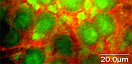
| Speaker | Dr. Peter J Verveer (Max Planck Institute of Molecular Physiology) |
| Title | Functional imaging of molecular
processes in living cells
|
| Abstract | In the last few decades, fluorescence microscopy has become a vertasile and indispensable tool for investigating molecular processes in living cells. It has become possible to measure the localization, activity and interactions of proteins in single living cells with high spatial and temporal resolution. In the first part of this lecture, the basics of fluorescence and of fluorescent labeling in cells will be discussed. In the second part the basics of fluorescence microscopy will be described, and recent developments in the field will be presented. In the last part, methods for measuring protein activity and interactions in living cells using fluorescence microscopy will be presented. |
| References | E. Haustein and P. Schwille. Fluorescence correlation spectroscopy: novel variations of an established technique. Annu. Rev. Biophys. Biomol. Struct. 36: 151-169, 2007. S. W. Hell. Far-field optical nanoscopy. Science 316:1153-1158, 2007. E. A. Jares-Erijman and T. M. Jovin. Imaging molecular interactions in living cells by FRET microscopy. Curr. Opin. Chem. Biol. 10:409-416, 2006. J. Lippincott-Schwartz and G. H. Patterson. Development and use of fluorescent protein markers in living cells. Science 300:87-91, 2003. F. S. Wouters, P. J. Verveer and P. I. H. Bastiaens. Imaging biochemistry inside cells. Trends Cell Biol. 11:203-211, 2001. |